A pressure ulcer is an ischemic lesion located in an area of the skin
and underlying tissue caused by sustained pressure / prolonged over two hard
planes, causing ischemia (produced by a number of forces that will be described
below) and necrosis.

The strengths responsible for the
onset of ulcers are:
- Pressure: force acting perpendicular to the skin as a result of gravity, that cause a tissue flattening between two hard planes, one belonging to the patient (heel, sacrum ..) and the other belonging to the outer (bed, chair, tube ..). A pressure above 32mmHg occludes capillary blood flow in soft tissue, causing hypoxia and therefore necrosis.
- Friction: tangential force that acts parallel to the skin, causing friction by movement or pull.
- Shear: combining friction and pressure. The shear force occurs when there is adjacent sliding surfaces (bed fowler position, what causes the patient to slide below).
In addition to the forces mentioned
above, are also involved:
- Maceration: produced by excessive moisture in the skin which leads to softening and reduced skin resistance.
- Poor nutritional status: hypoproteinemia, anemia, dehydration and vitamin deficiency affecting the integrity of tissues
The ulcers usually occur in partial
/ total bedridden patients or in patients who are sitting in one position a
long period of time. However, can also appear in patients with pressure
maintained in a defined area, such as the nose because of the pressure exerted
by an oxygen mask or on the ears or the pressure exerted by the rubber mask.
Typically, the ulcers are over bony prominences, being more sensitive (and
frequent) the shoulder blades, elbows, external malleolus, sacrum, coccyx,
heels, ischial tuberosities and trochanteric prominences.

Pathophysiological risk factors of occurrence of ulcers are skin lesions
(aging and related conditions), oxygen transport disorders, nutritional
deficiencies, altered state of consciousness (drugs, confusion and coma), motor
deficit (stroke, fractures , paralysis, paresthesia), sensory deficits (loss of
thermal and pain sensitivity), abnormal elimination (urine and feces).
Situational risk factors of occurrence of ulcers are: wrinkles in bed, poor hygiene, objects of
touch, pain, immobility,
The environmental risk factors of occurrence of ulcers are: misuse
prevention material, lack of unified criteria in planning cures, lack of
health education or malpractice of healthcare professionals.
When a patient presents with an ulcer, you have to make a comprehensive evaluation of this, taking into account:
- Background of the wound
- Personal history, family, and social drug.
- Physical examination of the patient and the wound
- Size
- Edema, erythema and warmth
- Wound bed (type of tissue, tendon exposure)
- Edges of the wound
- Characteristics of the wound edges (bagged, edema, pigmentation)
- Wound location, color, and odor exudate
- Temperature
- Blood pressure
- Neurological examination
- Arterial pulses
- Response to the elevation of the limb, and pain in the same
- Etiology of injury
- Diagnosis of comorbidities
- Current status of the wound
- Treatment Plan
The classification of ulcers is done
in 4 degrees / stages:
- Grade I: are those appearing cutaneous erythema (skin pink / red) that not giving up when you remove the pressure. In dark skin may appear purple. Only there is involvement of the epidermis.

- Grade II: are those in which it's produced a solution of continuity of the skin, vesicles and bullae. It affects the epidermis and the superficial dermis. Presents abrasion appearance or shallow crater.

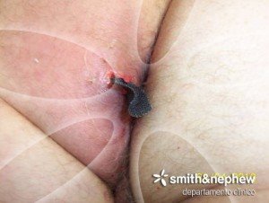
- Grade III: are those in which there is subcutaneous tissue affectation. The tissue necrosis extending deep through the skin, reaching the deep dermis and hypodermis. Lesions appear as deep crater (if not covered by necrotic tissue).

- Grade IV: are those in which there is a total loss of skin thickness and necrosis in deep structures (muscle, bone or supporting structures). Appear cavernous and tunneling lesions.

It is very important that, before
determining the degree of ulcer, removed the necrotic tissue in order to
properly assess the degree of involvement of deeper tissues.
For the assessment of the ulcers we
have the Norton scale, in which we make a valuation of: patient's general
physical condition, mental condition, activity, mobility and incontinence.

This scale must be done
continuously, as it may be modified by a variety of factors.
For the treatment of ulcers, it is
necessary to perform:
- Debridement: it's the remission of foreign material and devitalized (contaminated tissue) adjacent to a traumatic injury or contaminated until healthy tissue disappears. There are several types of debridement: surgical, autolytic, enzymatic, mechanical and others.

- Hydrocolloid dressings: these dressings decreased the oxygen tension and also reduce the pH in the ulcer (reduce the presence of bacteria). These dressings have the advantage that: they reduce infection rates, accelerate the healing process, causing less damage to the removal, autolytic, reduce odor and have better cost-effectiveness, provide comfort to the user.


- Alginates: derived from seaweed. Of these sodium alginate is extracted, which mecienta an exchange process with a solution containing calcium ions, produces precipitation of calcium alginate fibers, which are highly absorbent hemostatic products and biodegradable, which possess antibacterial activity.
- Hydrogels: contain lots of water. They are indicated in ulcers with minimal or moderate exudate. Have the feature that in addition to being occlusive, hydrate, relieve pain and they are debriding autolytic effective in surfaces with slough, bedsores and fibrin.

- Silver Antimicrobial Dressings: Silver prevents respiration and feeding bacterial, which inhibits bacterial enzymes and interferes with cellular respiration (the dressing is very useful, as the pus, necrotic tissue and slough are bacterial breeding grounds).
The prevention of pressure ulcers is
far more important than anything I mentioned above. It is essential to follow a
series of preventive measures such as:
- Mitigate or eliminate the pressure
- Frequent changes in position to avoid blocking blood flow
- Do not drag the patient to avoid friction
- Observe skin daily to see if there is redness or whitish areas
- Use decubitus, air or water mattresses
- Do not raise the head of the bed more than 30 degrees to prevent sliding pressure
- Make assets and liabilities exercise
- Dry thoroughly after bathing
- Bedding clean, dry and wrinkle
- Diet rich in proteins and vitamins (especially vitamin C)

In my opinion, it is vital to
prevent pressure ulcers, especially in patients immobilized because, once an
ulcer appears, has a long time to disappear.
Bibliography
- Álvarez C, Lorenzo M. Cuidados de enfermería en una población geriátrica con riesgos de úlcera por presión. Enfermería Global 2011; 23: 172-182. Disponible en: http://scielo.isciii.es/pdf/eg/v10n23/administracion3.pdf
- Martínez López, J. F. Prevención y tratamiento de úlceras y escaras. Málaga: Editorial vértice; 2008.
- Morales Martínez, F. Temas prácticos en geriatría y gerontología (Tomo 1). Costa Rica: Editorial Universidad Estatal a Distancia; 2007.
No hay comentarios:
Publicar un comentario